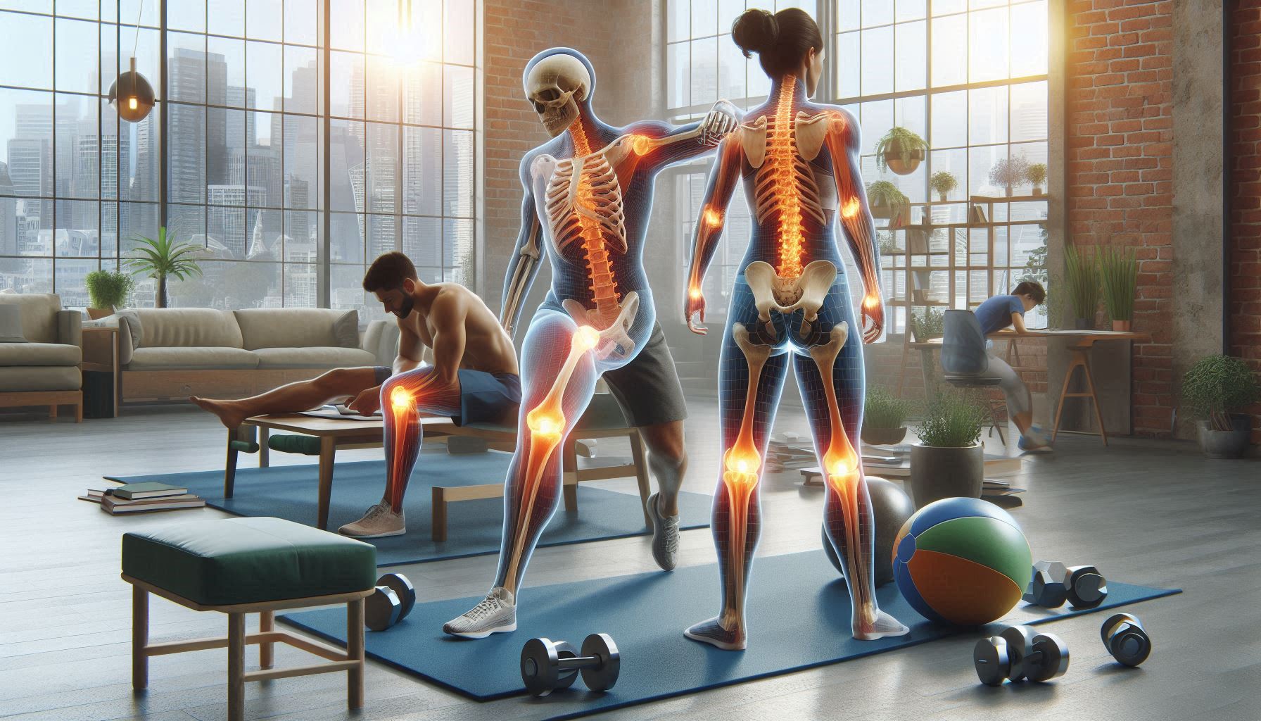
The human body is a marvel of engineering, a complex system of levers, pulleys, and shock absorbers designed for movement. At the core of this mobility are our joints—the hinges and ball-and-sockets that allow us to walk, run, reach, and lift. However, this constant use, compounded by injury, overuse, or the natural aging process, makes them susceptible to wear and tear. When a joint is compromised, the path to recovery is not a one-size-fits-all journey. The hips, shoulders, and knees, being three of the most critical and frequently injured joints, each demand a unique, nuanced approach to rehabilitation.
This guide provides a comprehensive exploration of the recovery processes for the hip, shoulder, and knee joints. We will dissect their unique anatomies, common ailments, and the specific, targeted strategies required to restore them to full function. Understanding these differences is the first step toward a successful and sustainable recovery.
The Hip Joint – Recovering the Foundation of Movement
Often called the body’s powerhouse, the hip is a classic ball-and-socket joint, designed for both immense stability and a significant degree of mobility. It bears the weight of the upper body and is central to virtually every lower body movement.
Anatomy and Biomechanics: The Engine Room
The femoral head (the “ball” of the femur) articulates with the acetabulum (the “socket” of the pelvis). This deep socket provides inherent stability. A complex network of ligaments and some of the body’s strongest muscles—the glutes, hamstrings, quadriceps, hip flexors, and deep lateral rotators—surround and control it. The health of the hip is also deeply connected to the core and the kinetic chain; weakness in the glutes or core can place abnormal stress on the hip joint and, subsequently, the knees and lower back.
Common Hip Injuries and Conditions
- Femoroacetabular Impingement (FAI): A condition where extra bone grows along one or both bones that form the hip joint, creating an irregular contour that causes friction and can damage the labrum and articular cartilage.
- Labral Tear: The labrum is a ring of cartilage that lines the acetabulum, providing stability and a suction seal. Tears can result from FAI, trauma, or repetitive motion.
- Osteoarthritis: The “wear-and-tear” arthritis that results from the breakdown of the protective articular cartilage covering the bones.
- Gluteal Tendinopathy (formerly Trochanteric Bursitis): Pain and dysfunction of the tendons of the gluteus medius and minimus muscles where they attach to the greater trochanter (the bony prominence on the side of the hip).
- Strains: Tears in the muscles or tendons surrounding the hip, commonly the hamstrings, hip flexors, or adductors (groin strain).
The Hip Recovery Protocol
Hip recovery prioritizes restoring stability without sacrificing necessary mobility. The focus is on activating the deep stabilizers and strengthening the primary movers.
Phase 1: Acute Phase (Protection & Pain Management)
- Goal: Reduce pain and inflammation, protect injured structures.
- Actions: Relative rest (avoiding painful movements), use of ice and NSAIDs as advised by a doctor, and potentially using crutches for a brief period if weight-bearing is painful.
- Early Mobilization: Gentle, pain-free range-of-motion exercises, such as supine heel slides and very gentle hip circumduction.
Phase 2: Subacute Phase (Restoring ROM & Early Strengthening)
- Goal: Regain full, pain-free range of motion and activate stabilizing muscles.
- Key Exercises:
- Neuromuscular Re-education: Clamshells, glute bridges, and side-lying leg lifts with a focus on consciously activating the gluteus medius and maximus, not the hamstrings or low back.
- Mobility: Gentle static stretching for the hip flexors, hamstrings, and piriformis if tightness is present. Quadruped rockbacks can help restore hip flexion.
- Isometric Strengthening: Gentle isometric holds in glute bridges or side-lying abduction.
Phase 3: Strengthening Phase (Building Resilience)
- Goal: Develop robust strength and endurance in the entire hip complex and core.
- Key Exercises: This phase moves into functional, weight-bearing movements.
- Monster Walks with a resistance band.
- Single-Leg Bridges and Single-Leg Deadlifts (bodyweight, then weighted).
- Lunges in all planes (forward, reverse, lateral).
- Step-Ups, focusing on controlled movement and glute activation at the top.
- Core Integration: Planks, Pallof presses, and dead bugs to ensure core stability supports hip function.
Phase 4: Return to Activity (Integration & Power)
- Goal: Safely reintegrate into sport-specific or high-impact activities.
- Key Exercises: Incorporating plyometrics and dynamic movements.
- Box Jumps and Jump Squats (progressing from double-leg to single-leg).
- Lateral Bounds and Skipping.
- Sport-Specific Drills: Gradual reintroduction of cutting, pivoting, and acceleration/deceleration patterns.
Prevention is Key: Maintaining strong glutes and a strong core is the best defense against hip injuries. Avoid prolonged sitting, which can lead to tight hip flexors and inactive glutes.
The Shoulder Joint – Rehabilitating Mobility and Control
The shoulder is the body’s most mobile joint, a trade-off that comes at the cost of inherent stability. This mobility is enabled by its structure: a ball-and-socket joint where the humeral head (the “ball”) is disproportionately larger than the glenoid fossa of the scapula (the “socket”). Stability, therefore, is provided dynamically by a complex system of muscles, tendons, and ligaments known as the rotator cuff and the scapular stabilizers.
Anatomy and Biomechanics: The Floating Platform
The shoulder is not one joint but four: the glenohumeral joint, the acromioclavicular (AC) joint, the sternoclavicular joint, and the scapulothoracic articulation. Proper function requires a perfect symphony between these parts. The rotator cuff muscles (supraspinatus, infraspinatus, teres minor, subscapularis) hold the humeral head centered in the shallow socket during movement. The scapula (shoulder blade) must move rhythmically and stably on the ribcage to provide a solid base for the arm to move upon. This concept is known as scapulohumeral rhythm.
Common Shoulder Injuries and Conditions
- Rotator Cuff Tendinopathy and Tears: Degeneration or tearing of the rotator cuff tendons, often from impingement, overuse, or acute trauma.
- Shoulder Impingement Syndrome: A condition where the rotator cuff tendons are compressed or “impinged” during arm elevation, often under the acromion bone.
- Labral Tears: The glenoid labrum can tear, most famously a SLAP tear (Superior Labrum from Anterior to Posterior), often seen in overhead athletes.
- Shoulder Instability and Dislocation: Often traumatic, where the humeral head is forced out of the socket, stretching or tearing the stabilizing ligaments (labrum and capsule).
- Frozen Shoulder (Adhesive Capsulitis): A condition characterized by stiffness, pain, and a significant loss of range of motion due to inflammation and thickening of the joint capsule.
The Shoulder Recovery Protocol
Shoulder rehab is a masterclass in balancing mobility and stability. The primary focus is almost always on strengthening the dynamic stabilizers—the rotator cuff and periscapular muscles—to recenter the joint and restore pain-free movement.
Phase 1: Acute Phase (Controlling Pain and Inflammation)
- Goal: Manage pain and protect healing tissues.
- Actions: Rest in a sling may be necessary post-surgery or dislocation. Modalities like ice and electrotherapy can help manage pain and inflammation. Pendulum exercises (Codman’s exercises) are introduced early to provide gentle distraction and mobilization.
Phase 2: Subacute Phase (Regaining ROM and Activating Stabilizers)
- Goal: Restore passive and active range of motion and begin retraining the rotator cuff and scapular muscles.
- Key Exercises:
- Range of Motion: Active-assisted range of motion (AAROM) using a stick or pulley system for flexion, abduction, and external rotation.
- Rotator Cuff Activation: Isometric internal and external rotation holds against a wall.
- Scapular Stabilization: The foundation of shoulder health. Exercises include scapular retraction (squeezing shoulder blades together without shrugging), scapular punches in a plank position, and wall slides.
Phase 3: Strengthening Phase (Building a Stable Base)
- Goal: Develop strength and endurance in the dynamic stabilizers and integrate shoulder function with the core.
- Key Exercises: This phase uses light resistance (bands, light dumbbells) with high repetitions.
- Rotator Cuff Strengthening: External and internal rotation with resistance bands, “Empty Can” exercises for supraspinatus (with caution).
- Scapular Strengthening: Rows, prone Y-T-W-L exercises, and serratus anterior punches (push-up plus).
- Integrated Movements: Overhead press and pulling motions are reintroduced very gradually, ensuring proper scapular movement is maintained.
Phase 4: Return to Activity (Sport-Specific Training)
- Goal: Prepare the shoulder for the demands of specific sports or occupations (e.g., throwing, swimming, lifting overhead).
- Key Exercises:
- Plyometrics: Medicine ball chest passes, overhead throws.
- Advanced Stabilization: Push-ups on unstable surfaces, bodyweight arcs.
- Graduated Throwing Program: For overhead athletes, a meticulously planned program that slowly increases throwing distance and intensity.
Prevention is Key: The best prevention for shoulder injuries is maintaining excellent posture and a consistent regimen of rotator cuff and scapular strengthening exercises, especially for overhead athletes and desk workers.
The Knee Joint – Restoring Stability and Load-Bearing Capacity
The knee is a modified hinge joint, primarily designed for flexion and extension (bending and straightening), with a small amount of rotation. Its primary role is to absorb shock and transmit force during weight-bearing activities. It is the largest joint in the body and, consequently, one of the most commonly injured.
Anatomy and Biomechanics: The Complex Hinge
The knee joint is formed by the femur (thigh bone), tibia (shin bone), and patella (kneecap). Its stability comes not from a deep bony socket but from a sophisticated system of ligaments and cartilage:
- Ligaments: The Anterior Cruciate Ligament (ACL) and Posterior Cruciate Ligament (PCL) control forward and backward translation of the tibia. The Medial Collateral Ligament (MCL) and Lateral Collateral Ligament (LCL) control side-to-side stability.
- Meniscus: Two C-shaped wedges of cartilage (medial and lateral) that act as shock absorbers between the femur and tibia, distributing load and providing stability.
- Muscles: The quadriceps (front of thigh) and hamstrings (back of thigh) are the primary movers and dynamic stabilizers of the knee. The glutes and calves also play a critical role in knee mechanics.
Common Knee Injuries and Conditions
- ACL Tear: A rupture of the anterior cruciate ligament, often a non-contact injury involving cutting, pivoting, or landing awkwardly.
- Meniscal Tear: A tear in the shock-absorbing meniscus, which can be traumatic (twisting) or degenerative.
- Patellofemoral Pain Syndrome (PFPS): “Runner’s knee,” characterized by pain behind or around the kneecap, often due to maltracking caused by muscle imbalances.
- Osteoarthritis: Degeneration of the articular cartilage on the surfaces of the femur, tibia, and/or patella.
- Tendinopathies: Such as patellar tendinitis (“jumper’s knee”) or quadriceps tendinitis.
The Knee Recovery Protocol
Knee rehabilitation is heavily focused on restoring stability, proprioception (joint position sense), and the strength of the muscles that dynamically control the joint, particularly the quadriceps, hamstrings, and glutes.
Phase 1: Acute Phase (Reducing Swelling and Restoring ROM)
- Goal: Control effusion (swelling), manage pain, and regain full knee extension.
- Actions: The RICE protocol (Rest, Ice, Compression, Elevation) is critical. Restoring full extension is a primary early goal, often achieved through heel props and gentle passive stretching. Early quadriceps activation is also paramount.
Phase 2: Subacute Phase (Building a Foundation of Strength)
- Goal: Restore normal gait (walking), improve strength, and begin neuromuscular training.
- Key Exercises:
- Quadriceps Activation: Straight leg raises, isometric quad sets, and mini squats.
- Hamstring and Glute Activation: Hamstring curls (lying down), glute bridges, and hip thrusts.
- Proprioception/Balance: Weight-shifting drills, single-leg stance, and eventually balance on an unstable surface (e.g., foam pad).
Phase 3: Strengthening Phase (Developing Robustness)
- Goal: Build maximal strength, single-leg strength, and control to handle higher loads.
- Key Exercises: This phase is heavily focused on closed-chain (foot on the ground) exercises.
- Double-Leg to Single-Leg: Progressing from squats and leg presses to split squats, Bulgarian split squats, and single-leg squats.
- Eccentric Control: Emphasis on the lowering phase of exercises, which is crucial for tendon health and injury prevention (e.g., eccentric squats, Nordic hamstring curls).
- Plyometric Preparation: Introduction of low-impact jumping and landing drills, focusing on “soft” landings with bent knees and proper hip hinge.
Phase 4: Return to Sport (Power and Agility)
- Goal: Safely return to cutting, pivoting, and jumping sports.
- Key Exercises:
- Plyometrics: Box jumps, jump squats, lateral hops, and single-leg hops for distance.
- Agility Drills: Ladder drills, cone drills, and sport-specific change of direction patterns.
- Gradual Exposure: A structured program that slowly reintroduces the specific stresses of the individual’s sport, ensuring the knee is prepared for unpredictability.
Prevention is Key: Knee injury prevention programs (like the FIFA 11+) that focus on strengthening the hamstrings and glutes, improving landing mechanics, and enhancing proprioception have been proven to significantly reduce the risk of ACL and other knee injuries.
Conclusion
Successful rehabilitation for any major joint, whether hip, shoulder, or knee, is built upon a common, non-negotiable foundation: an accurate diagnosis to treat the root cause, not just the symptoms. This journey demands patience and consistency, as tissue healing follows a biological timeline that cannot be rushed. It is crucial to listen to your body, distinguishing between “good pain” of muscle fatigue and the “bad pain” that signals harm, using this feedback to guide exercise modification. Recovery hinges on rebuilding the mind-muscle connection for neuromuscular control and stability through simple, focused exercises, while also addressing the entire kinetic chain understanding that a knee issue may stem from weak hips or a shoulder problem from a stiff spine. By adhering to this phased, principles-based approach, you can relearn your body’s mechanics, often emerging from injury stronger, more resilient, and more in tune with your body than before.
SOURCES
Biel, A., & Dorn, R. (2019). Trail Guide to the Body: A hands-on guide to locating muscles, bones and more (6th ed.). Books of Discovery.
Cook, G., Burton, L., Hoogenboom, B. J., & Voight, M. (2014). Functional movement screening: the use of fundamental movements as an assessment of function – part 1. International Journal of Sports Physical Therapy, 9(3), 396–409.
Hewett, T. E., Myer, G. D., & Ford, K. R. (2005). Reducing knee and anterior cruciate ligament injuries among female athletes: a systematic review of neuromuscular training interventions. Journal of Knee Surgery, 18(1), 82–88.
Lewis, J. (2009). Rotator cuff tendinopathy: a model for the continuum of pathology and related management. British Journal of Sports Medicine, 44(13), 918–923.
Manske, R., & Reiman, M. (2013). Non-operative management of femoroacetabular impingement: A prospective, randomized trial. Journal of Orthopaedic & Sports Physical Therapy, 43(11), 872-879.
Page, P. (2012). Current concepts in muscle stretching for exercise and rehabilitation. International Journal of Sports Physical Therapy, 7(1), 109–119.
Reiman, M. P., & Lorenz, D. S. (2011). Integration of strength and conditioning principles into a rehabilitation program. International Journal of Sports Physical Therapy, 6(3), 241–253.
Silvers, H. J., & Mandelbaum, B. R. (2011). Prevention of anterior cruciate ligament injury in the female athlete. British Journal of Sports Medicine, 41(1), 52–59.
Wilk, K. E., Obma, P., Simpson, C. D., Cain, E. L., Dugas, J. R., & Andrews, J. R. (2009). Shoulder injuries in the overhead athlete. Journal of Orthopaedic & Sports Physical Therapy, 39(2), 38–54.
HISTORY
Current Version
Aug 23, 2025
Written By:
SUMMIYAH MAHMOOD